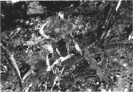
Figure 36.
Fibrous serpentine vein (S) cut into sections by late veins of calcite (C) in the serpentinized peridotite (Interval 149-897D-13R-1, 73-76 cm). Crossed-plane polarized light. The longer dimension of the photomicrograph represents 1.2 mm.