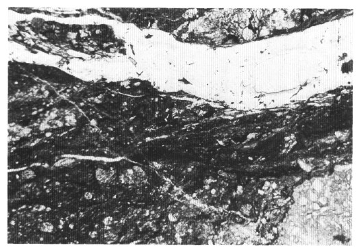
Figure 38. Localized shear zone displaying grain-size reduction, C-S fabric and rotated clasts at the boundary of a fragmented clast (Sample 149-899B-21R-4, 43 cm). Photomicrograph under cross-polarized light, the width of the vein in the upper part of the photo is on the order of 1 cm across.