![]()
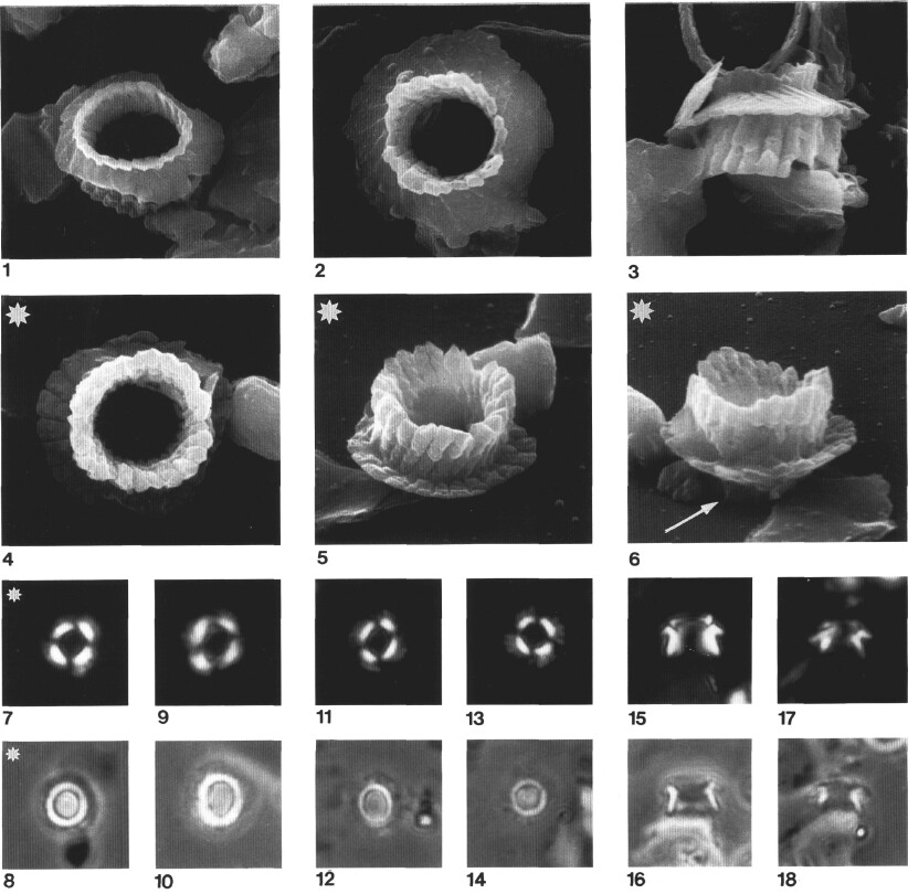
Plate 1. The illustrations of the Plates 1-10 are scanning electron (SEM) and light microscope (LM) micrographs. XP = cross-polarized light; Ph = phase contrast. Transferred specimens from the LM to the SEM are denoted on the plate captions by the superscript. Magnification for all LM micrographs is X2500 and magnification for SEM micrographs as indicated (X6000, X8000, or X12,000). Holotype specimens are indicated by an asterisk. Ansulasphaera covingtonii sp. nov.; all SEM specimens from Sample 149-901A-SR-1, 31 cm. 1. Oblique distal view, FSU-F4. 2. Distal view, FSU-F118. 3. Lateral view, FSU-F3. 4-8TR. Holotype, same specimen. (4) Proximal view, FSU-F68. (5) Oblique proximal view, FSU-F69. (6) Oblique proximal view, turned 180° as compared to Figure 5, FSU-F70. (7) XP, FSU-FO51-D26. (8) Ph, FSU-FO51-D25. 9, 10. 149-901A-3R-1, 2 cm; XP, FSU-FO41-D35 and Ph, FSU-FO41-D36. 11, 12. 149-901A-5R-1, 31 cm; XP, FSU-FO44-D4 and Ph, FSU-FO44-D5. 13, 14. 149-901A-5R-1, 31 cm; XP, FSU-FO45-D3 and Ph, FSU-FO45-D4. 15, 16. 149-901A-5R-1, 31 cm; XP, FSU-FO46-D13 and Ph, FSU-FO46-D14. 17, 18. 149-901A-5R-1, 31 cm; XP, FSU-FO45-D18 and Ph, FSU-FO45-D19. Magnification for all SEM micrographs is X8000.
![]()