![]()
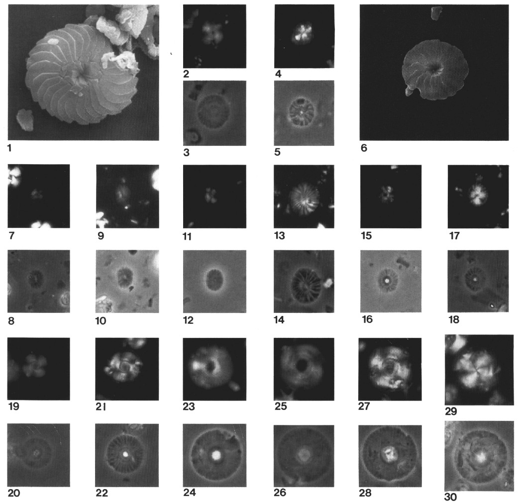
Plate 1. The illustrations
of Plates 1-14 are scanning electron microscope (SEM) and light microscope (LM)
micrographs. XP = cross-polarized light; Ph = phase contrast; PL =
parallel light. Transferred specimens from the LM to the SEM are denoted on the
plate captions by TR. Magnification for all LM micrographs is ×1700,
except where otherwise indicated, and magnification for SEM micrographs is
indicated (×3000, ×4000, ×5000, ×6000, or ×8000). Each plate represents one
major group of forms, which may include one or several genera. 1-6. Calcidiscus
leptoporus (Murray and Blackman) Loeblich and Tappan. (1, 6) Distal view, ×6000,
Sample 149-900A-22R-1, 122 cm. (2, 3) Sample 149-900A-25R-1, 79 cm, XP (2) and
Ph (3). (4, 5) Sample 149-900A-34R-2, 83 cm, XP (4) and Ph (5). 7-10. Calcidiscus
fuscus (Backman) Janin. (7, 8) Sample 149-900A-32R-3, 7 cm, XP (7) and Ph
(8). (9, 10) Sample 149-900A-38R-2, 57 cm, XP (9) and Ph (10). 11, 12. Calcidiscus
pataecus (Gartner) n. comb., Sample 149-900A-29R-1, 122 cm, XP (11) and Ph
(12). 13, 14. Calcidiscus radiatus (Kamptner) Martin Perez and Aguado,
Sample 149-900A-32R-2, 100 cm, XP (13) and Ph (14). 15-18. Calcidiscus
tropicus (<6.0 µm) (Kamptner) Varol. (15, 16) Sample 149-900A-30R-4, 16
cm, XP (15) and Ph (16). (17, 18) Sample 149-900A-30R-2, 41 cm, XP (17) and Ph
(18). 19, 20. Calcidiscus carlae (Lehotayova and Priewalder) Janin,
Sample 149-900A-12R-4, 10 cm, XP (19) and Ph (20). 21-28. Calcidiscus
tropicus (>6.0 µm) (Kamptner) Varol. (21, 22) Sample 149-900A-24R-5, 44
cm, XP (21) and Ph (22). (23, 24) Sample 149-900A-16R-1, 133 cm, XP (23) and Ph
(24). (25, 26) Sample 149-900A-14R-5, 142 cm, XP (25) and Ph (26). (27, 28) XP,
Sample 149-900A-15R-1, 16 cm, XP (27) and Ph (28). 29, 30. Calcidiscus
macintyrei (Bukry and Bramlette) Loeblich and Tappan, Sample 149-900A-12R-1,
48 cm, XP (29) and Ph (30).
![]()