![]()
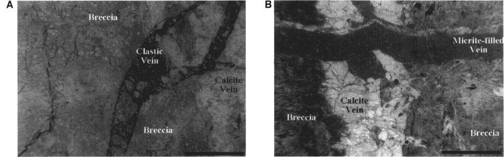
Figure 3. Two micrite-filled veins displaying clastic texture from Site 897. A. Plane-polarized light image of a highly oxidized vein in Sample 149-897D-13R-5, 57-61 cm, containing fragments of brecciated serpentinite. The clastic vein is observed to cut a calcite vein at the right side of the image. Scale bar is 5 mm long. B. Plane-polarized light image of a relatively homogeneous micrite-filled vein in Sample 149-897C-64R-4, 16-19 cm, which cuts across a large vein grading from vacuolized botryoidal calcite at the margins to clear, sparry calcite in the center. Clear, fibrous to sparry calcite is observed to postdate the emplacement of the micrite-filled vein; a thin vein parallels the top edge of the clastic vein and crosscuts a protrusion. Several perpendicular fibrous veins also cut the micrite-filled vein to the right of its intersection with the large vein. Scale bar is 5 mm long.
![]()