![]()
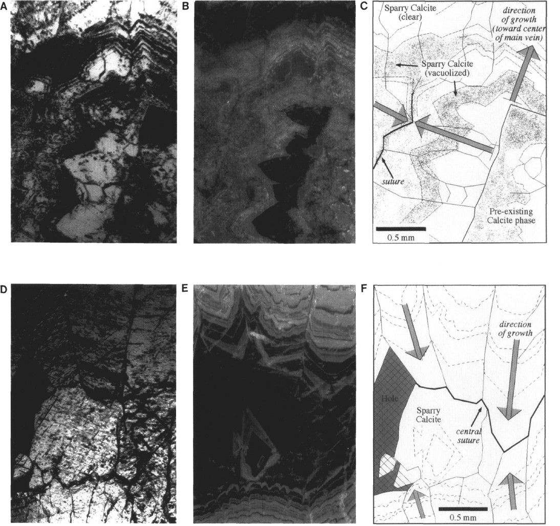
Figure 6. Sparry calcite vein filling, common to zoned veins at both Sites 897 and 899, from Sample 149-899B-18R-2, 135-139 cm. A. Plane-polarized light photomicrograph of inclusion-rich domain near the vein wall (not shown, but to lower right of photograph), showing complicated sawtooth growth patterns and inclusion banding at intersection with secondary zoned vein; calcite crystals initially grew outward from the vein walls and a preexisting calcite phase, simultaneously filling the secondary vein (at left) and main vein (at top). Once the secondary vein was sealed, crystal growth continued in the main vein. B. Cathodoluminescence photomicrograph of same view, showing high- to low-luminescent zones corresponding to bands of inclusions. C. Interpretive sketch of same view. Arrows show directions of mineral growth; dashed form lines show luminescent zoning patterns. D. Plane-polarized light photomicrograph of clear, coarse, bladed calcite near the center of the vein; grain boundaries stand out in relief, but are not otherwise distinguished (black hole to left is open porosity). E. Cathodoluminescence photomicrograph of same view, showing fine, rhythmic brightness zoning, grading to nonluminescent calcite near the center of vein. F. Interpretive sketch of same view, showing directions of growth, and position of central suture (symbols the same as in C).
![]()