![]()
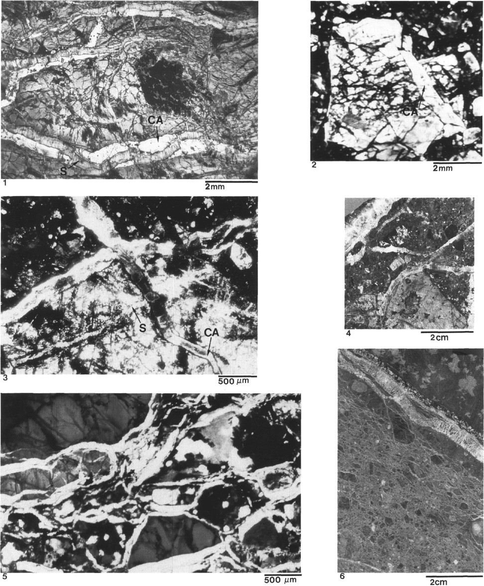
Plate 2. Calcite (deformational phase F3) and serpentine veins (deformational stage D) from Hole 899B. 1. Sample 149-899B-31R-1, 67-71 cm. Thin-section photomicrograph of serpentinite (S) veins overgrown by calcite (CA) within a clast of serpentinized peridotite. Plane-polarized light. 2. Sample 149-899B-19R-1,6-11 cm. Thin-section photomicrograph of a chrysotile vein (CR) in a serpentine-magnetite clast included within a cataclastic breccia. Plane-polarized light. 3. Sample 149-899B-18R-4, 99-110 cm. Thin-section photomicrograph of calcite veins (CA) that cross-cut the cataclastic breccia. Note calcite veins surrounding and cross-cutting large porphyroclast including serpentine veins (S). Crossed nicols. 4. Sample 149-899B-18R-2, 39-44 cm. Hand-specimen photograph showing calcite veins that cross-cut porphyroclast and matrix. Note the absence of shear movement associated with calcite veins; note also the large "dipping" vein with antiaxial filling in the upper left corner. 5. Sample 149-899B-16R-2, 23-27 cm. Thin-section photomicrograph showing complex network of calcite veins bounding angular porphyroclast and breccia fragments (jigsaw-puzzle pattern). 6. Sample 149-899B-21R-2, 40-55 cm. Mesoscopic jigsaw-puzzle pattern network of calcite veins (similar to (5)) in breccia. In the upper right corner is a large calcite vein, with an antiaxial filling, bounding a relatively fresh peridotite clast. Note lateral branches of the large vein merging with the network of minute veins bounding clasts. Note weak cataclastic foliation indicative of shear between peridotite clast and breccia.
![]()