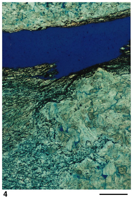
Plate 3. Photomicrographs. 4. Sample split along matrix-rich seam producing large epoxy-filled fracture seen in upper part of photograph. Fine lamination and detrital grains within the matrix-rich drape (left) interfinger and cover coarse, interlocking gypsum crystals in pinch-and-swell structure (right). For clarification, see core photograph in
Figure 7. Section 975B-34X-1, 26-30 cm. Scale bar is 0.2 mm.