![]()
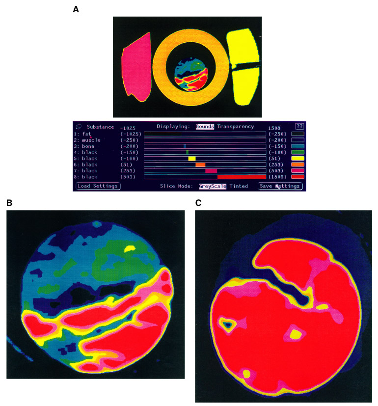
Figure 4. X-ray CT imagery showing (A) definite differences in color among methane hydrates (dark blue, turqouise, green), minerals (red, yellow), water ice (yellow), dry ice (pink) and acrylle tube (orange); (B) nodular methane hydrates contained in sediments; and (C) vein hydrates (dark blue) penetrating sediments (red) that are coincident with flaky hydrate.
![]()