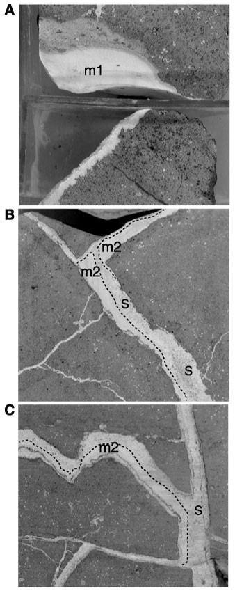
Figure 4. Macroscopic fracture-filling carbonate phases. A. Lens of fine-grained carbonate sediment, micrite 1 (m1), composed of recrystallized calcite most likely derived from nannoplankton fragments. The orientation of the sample is not preserved, but it is likely that this was deposited gravitationally at the bottom of the cavity (interval 165-1001A-54R-5, 13-18 cm). B. Fracture filled by micrite 2 (m2) and sparry calcite (s). Note the upper boundary of micrite 2, which suggests that lithification took place before further enlargement of the fracture and precipitation of sparry calcite 2 (s) (interval 165-1001A-54R-2, 63-68 cm). C. Fracture partially filled by micrite 2 with a geopetal orientation at the lower side of the fracture, and later by sparry calcite (s) (interval 165-1001A-54R-2, 51.5-55 cm).
![]()