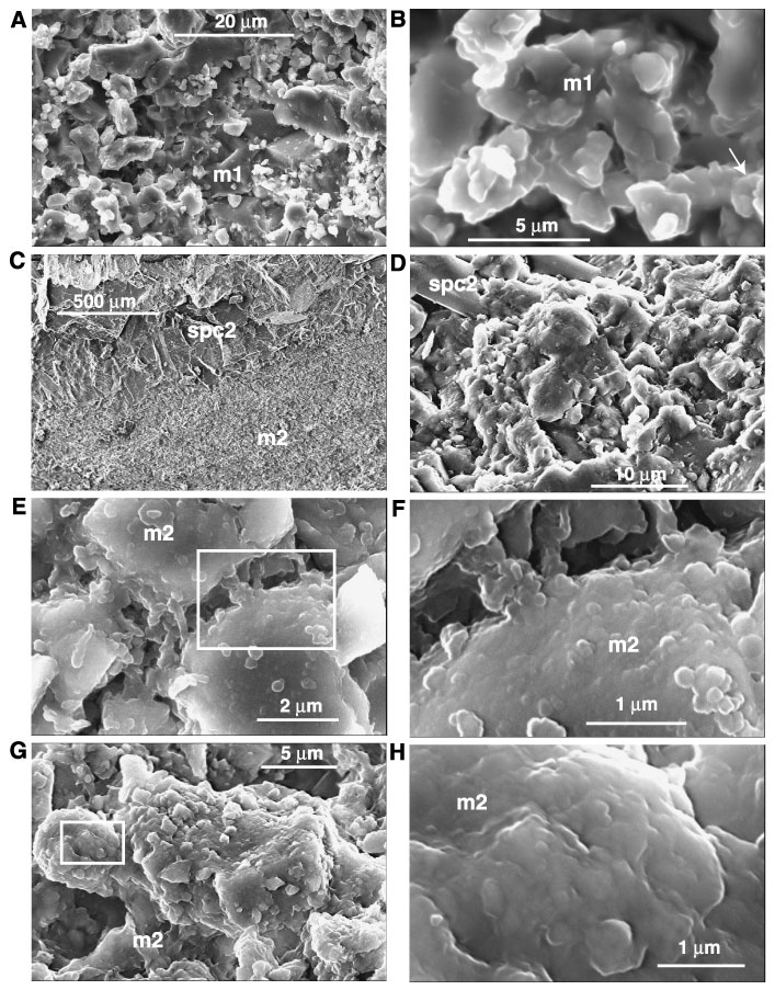
Figure 6. SEM photomicrographs. A. General aspect of micrite 1 (m1). Note the smooth surfaces of the crystals (interval 165-1001A-54R-5, 13-18 cm; see Fig. 3A). B. Close-up image of micrite 1 where it is possible to recognize the remnants of nannofossils (arrow) now almost entirely obliterated by recrystallization (interval 165-1001A-54R-5, 13-18 cm). C. Contact between micrite 2 (m2) and sparry calcite 2 (spc2) (interval 165-1001A-54R-1, 72-74 cm). D. Close-up of C, showing the detailed relationships between micrite 2 and sparry calcite 2 (spc2). Note the difference in appearance between micrite 1 as shown in A, and micrite 2 shown here. The crystals forming micrite 2 here are anhedral and their surfaces have a blobby appearance supporting microbial influence (interval 165-1001A-54R-1, 72-74 cm). E. View of micrite 2, made of euhedral to subhedral crystals with a blobby surface. The white rectangle indicates the field of view of image F (interval 165-1001A-54R-1, 72-74 cm). F. Detailed view of image E showing bridges between grains supporting microbial influence (interval 165-1001A-54R-1, 72-74 cm). G. The white rectangle indicates the field of view of image H (interval 165-1001A-54R-4, 67-72 cm). H. Blobby crystal surface forming a cauliflower structure supporting microbial influence (interval 165-1001A-54R-4, 67-72 cm).
![]()