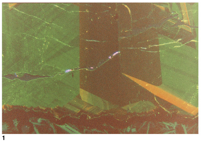
Plate 1. Photomicrographs of carbonate veins from Leg 168 basalt specimens, taken using cathodoluminescence (CL). 1. Blocky aragonite vein with basalt host rock visible along bottom edge. The aragonite is optically clear and homogeneous; however, the CL image reveals complex zones of varying CL color and intensity. Purple luminescence is from polishing compound (alumina?) stuck in epoxy that fills a microcrack through the section. Sample 168-1027C-5R-4, Piece 9.
![]()