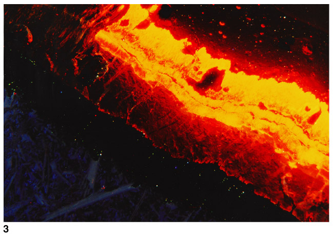
Plate 1. Photomicrographs of carbonate veins from Leg 168 basalt specimens, taken using cathodoluminescence (CL). 3. Zoned vein with cross-fiber calcite in the middle (bright CL) and blocky aragonite (dark CL) at the edge. Host rock is visible at lower left. Sample 168-1032A-12R-1, Piece 1.
![]()