![]()
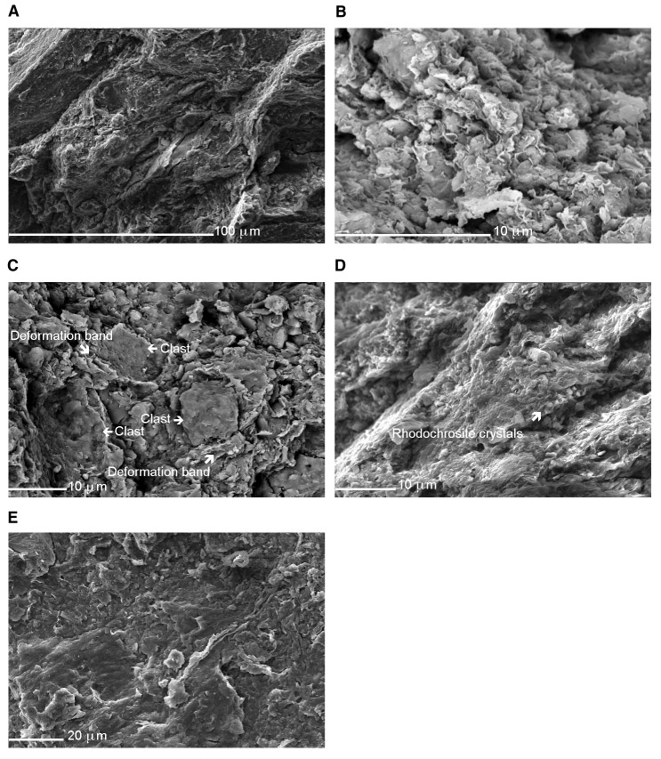
Figure F8. Sequence of secondary electron micrographs, showing relevant features of the décollement. A. Fracture arrays in the upper part dividing the material into blocky fragments. B. Undeformed clays in between fractures. C. Draping of clay minerals around siltier grains. D. Fracture surfaces, localizing in clay-rich domains. E. Looking flat on to a shear surface. Note the planar nature and intense grain alignment.
![]()