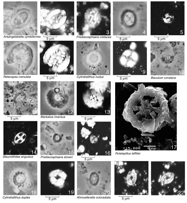
Plate P3. Magnification for light micrography is 2200x. Light micrography: PH = phase-contrast light, PL = plain transmitted light, XP = cross-polarized light, SEM = scanning electron micrography. 1, 2. Arkhangelskiella cymbiformis, Sample 173-1068A-15R-3, 71-73 cm, PH (1) and XP (2). 3, 4. Prediscosphaera cretacea, Sample 173-1068A-15R-3, 34-35 cm, PH (3) and XP (4). 5, 10. Biscutum constans, Sample 173-1069A-12R-3, 27-29 cm, XP (5) and PH (10). 6, 7. Retecapsa crenulata, Sample 173-1068A-15R-2, 92-93 cm, PH (6) and XP (7). 8, 9. Cylindralithus nudus, Sample 173-1069A-12R-4, 4-6 cm, PH (8) and XP (9). 11, 14. Staurolithites angustus, Sample 173-1069A-12R-3, 27-29 cm, PH (11) and XP (14). 12, 13. Markalius inversus, Sample 173-1068A-14R-1, 6-8 cm, PH (12) and XP (13). 15, 16. Prediscosphaera stoveri, Sample 173-1069A-12R-3, 134-136 cm, PH (15) and XP (16). 17. Rotelapillus laffittei, Sample 173-1069A-15R-2, 71-75 cm, SEM, magnification indicated. 18, 19. Cylindralithus duplex, Sample 173-1069A-12R-2, 99-100 cm, PH (18) and XP (19). 20-22. Ahmuellerella octoradiata, Sample 173-1068A-14R-7, 35-36 cm, PH (20) and XP (21, 22). Click on image for close-up.
![]()