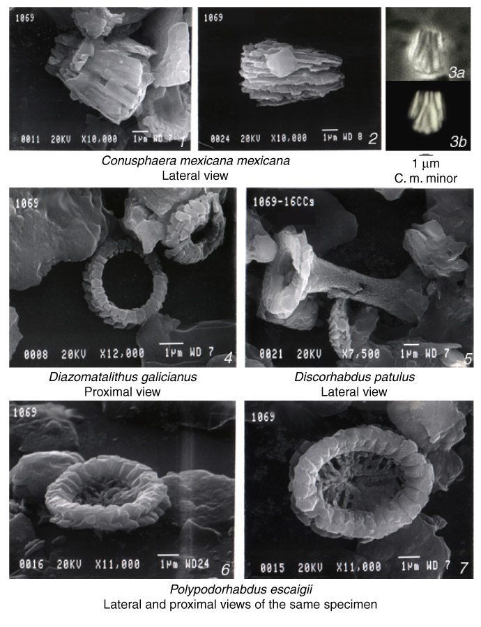
Plate P2. Scanning electron micrographs of calcareous nannofossils from Sample 173-1069A-16R-3, 129 cm, except for figure 3. 1, 2. Partially etched specimens showing inner cores of the conusphaerids. 3. (a) phase-contrast and (b) cross-polarized light micrographs of a specimen from Sample 173-1065A-11-CC, 14-16 cm. (figs. 2, 4, and 5 are from Wilson et al., in press.) Click on image for close-up.
![]()