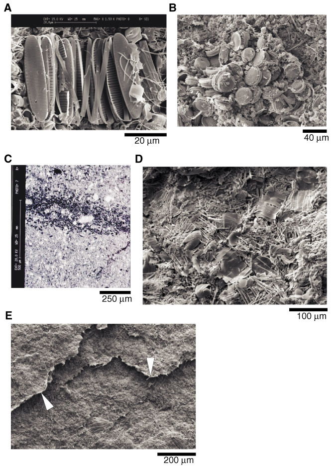
Figure F5. A. SEI micrograph of a cluster, or intact colony, of Fragilariopsis pennate diatoms within a diatom-ooze lamina (Sample 178-1098A-6H-1, 54.1 cm). B. SEI micrograph of a cluster of Thalassiosira antarctica within the very top of a diatom-bearing terrigenous lamina, possibly a remnant of a fecal pellet (Sample 178-1098A-6H-1, 52.1 cm). C. BSEI micrograph through a centric diatom sublamina within a diatom-bearing terrigenous lamina. The darker, lower backscatter coefficient diatom sublamina is ~250 µm thick (Sample 178-1098A-6H-3, 4.5-8 cm). D. SEI micrograph of Corethron criophilum sublamina from the mid-to-base region of a diatom-bearing terrigenous lamina (Sample 178-1098A-6H-1, 53.8 cm). E. SEI micrograph of a lamina-parallel fracture surface through a diatom-bearing terrigenous lamina. Frayed edges of Chaetoceroetae sublaminae are indicated by arrows (Sample 178-1098A-6H-1, 53.6 cm).
![]()