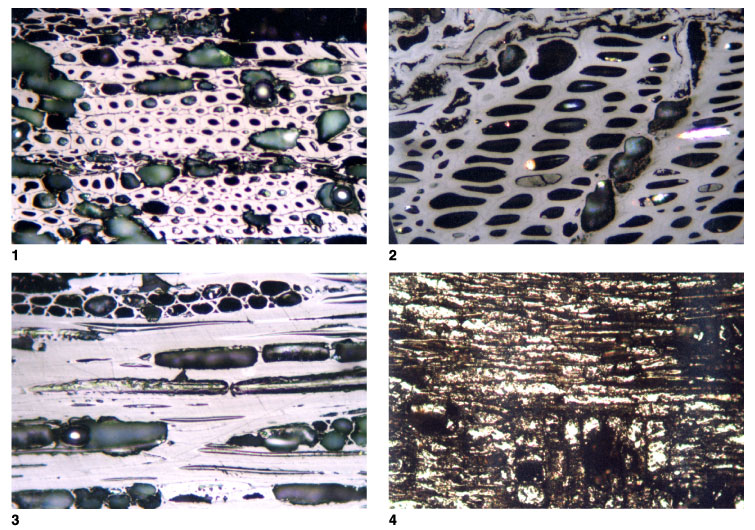
Plate P8.
Pyrofusinite and pyrite replacement of woods. 1.
T5829; Hole 1115A; 272.84 mbsf. Fusinite formed by charring of woody tissues.
Fusinite is generally absent from the sample suite but is dominant in two
samples. In these samples, probable air-fall tuff fragments are present, but
such tuff fragments are present in many samples lacking fusinite. Reflectance of
secondary cell walls = 3.46%. Primary cell walls have a much lower reflectance
but are too thin to measure accurately (reflected light; field width = 0.22 mm;
mean vitrinite reflectance = 0.22% and 0.25% in adjacent samples). 2.
T5859; Hole 1118C; 725.88 mbsf. Material close to the fusinite to semifusinite
boundary but formed by the charring of woody tissues. Some commercially
available artificial chars show reflectance down to 0.7%. Reflectance of
secondary cell walls = 1.10%. As for figure 1, the primary cell walls have a
lower reflectance than the secondary walls (reflected light; field width = 0.22
mm; mean vitrinite reflectance = 0.33% and 0.28% in adjacent samples). 3.
T5829; Hole 1115A; 272.84 mbsf. Fusinite formed by charring of woody tissues, in
this case xylem tissues, seen in longitudinal section. Cell wall structures are
preserved, and some probable pit structures, probably scalariform pitting, are
seen in some of the cell lumens. Reflectance of secondary cell walls = 3.96%.
Primary cell walls seem to be similar to the secondary cell walls in terms of
reflectances in this field (reflected light; field width = 0.22 mm; mean
vitrinite reflectance = 0.22% and 0.25% in adjacent samples). 4.
T5840; Hole 1115C; 489.50 mbsf. Pyrite petrifaction of wood tissue. The darker
areas are cell walls preserved as vitrinite. The tissue is xylem seen in
longitudinal section, and the vertical structures are medullary rays (reflected
light; field width = 0.22 mm; mean vitrinite reflectance = 0.29%). Click on
image or number to see enlargement.