Plate P3.
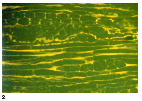
Vitrinite showing fluorescence from cell walls. 2. T5809; Hole
1109D; 387.86 mbsf. Same as figure 1, but in fluorescence mode. Longitudinal
section of wood with the cells outlined by the fluorescence of the cell walls.
The structures present are probably xylem, seen in longitudinal section, with
some medullary ray tissues in the upper part of the field. Fluorescence of the
cell walls is similar to that for suberinite, but the form of the cells shows
that this material is not from cork cells and cannot strictly be referred to the
maceral suberinite (reflected light; field width = 0.22 mm; vitrinite
reflectance [cell contents] = 0.37%, [cell walls] = 0.12%).