![]()
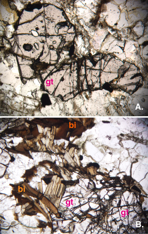
Figure F46. Photomicrographs of garnet gneiss. A. Poikiloblastic garnet (gt) in Unit 6 (clast in conglomerate, Sample 183-1137A-35R-2, 46-47 cm). Field of view = 1.4 mm (plane-polarized light). B. Porphyroblastic garnet (gt) and biotite (bi) from Unit 9 (clast in tuff, Sample 183-1137A-44R-4, 44-46 cm). Field of view = 2.8 mm (plane-polarized light).
![]()