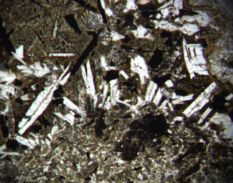
Figure F21. Margin of a segregation showing plagioclase crystals that nucleated at the contact and grew toward the center of the segregation, which is at the top of the image. Sample 183-1136A-17R-2 (Piece 6, 57-60 cm) in plane-polarized light; width of field of view = 2 mm.
![]()