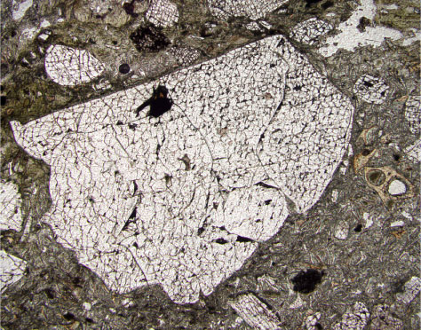
Figure F51. Photomicrograph of a fractured sanidine phenocryst from Unit 9 tuff (Sample 183-1137A-44R-4, 6-9 cm). The outlines of glass shards in the matrix can be seen. The image is in plane-polarized light; field of view width = 2.75 mm.