![]()
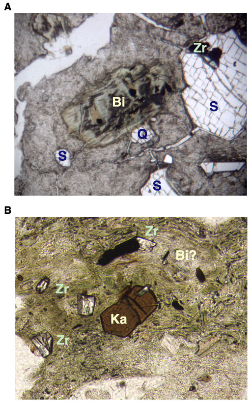
Figure F53. Photomicrographs highlighting the breakdown of mafic crystals in the Unit 9 tuff (Sample 183-1137A-41R-3, 42-44 cm). A. Original biotite (Bi) has been replaced by relatively well-crystallized clay; inclusions of titanomagnetite, zircon (Zr), and kaersutite are common. A quartz crystal (Q) is just below the altered biotite; note titanomagnetite and zircon in the large sanidine crystal (S) in the upper right hand corner. The image is in plane-polarized light; field of view width = 2.75 mm. B. Isolated kaersutite (Ka), zircon, and titanomagnetite, and perhaps a relic biotite (Bi). The image is in plane-polarized light; field of view width = 1.4 mm.
![]()