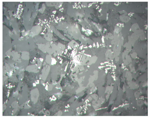
Figure F58. Close-up photomicrograph of skeletal, dendritic titanomagnetite groundmass crystals in the flow margin of Unit 19. Sample 183-1138-87R-2, 110-113 cm, in reflected light; width of field of view = 0.5 mm.