Figure
F71. Color
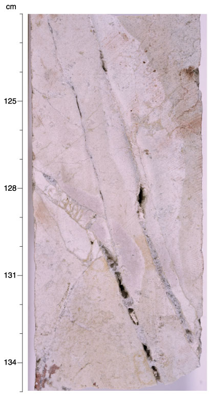
close-up photograph of Sample 183-1139A-70R-2 (Piece 1, 122-135 cm). Completely
altered white to very pale green to light pink sanidine-phyric trachyandesite.
Most intense alteration (white and pink) occurs as halos along veins filled with
quartz, siderite, and calcite. Sanidine, siderite, and quartz were the only
phases identified by XRD analysis of the white wall rock. Rock is locally
brecciated between veins.