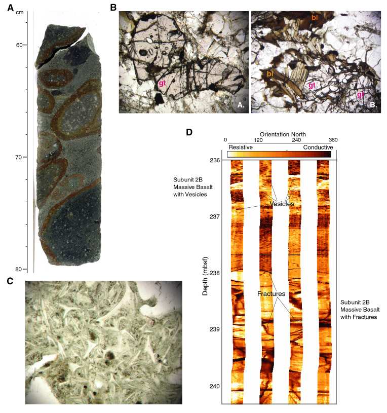
Frontispiece. A. Core photo of conglomerate from Site 1137 containing rare clasts of garnet-biotite gneiss. Interval 183-1137A-34R-3, 57–80 cm. B. Photomicrographs of garnet gneiss clasts from conglomerate (left) and crystal-vitric tuff (right) at Site 1137 showing poikiloblastic garnet (gt) and biotite (bt). Left: Sample 183-1137A-35R-2, 46–47 cm; field of view = 1.4 mm. Right: Sample 183-1137A-44R-4, 44–46 cm; field of view = 2.8 mm. C. Photomicrograph of crystal-vitric tuff at Site 1137 showing well-preserved cuspate and tricuspate glass shards, which form during explosive fragmentation of vesiculating magma. Sample 183-1137A-44R-4, 44–46 cm. Field of view = 1.40 mm. D. Formation MicroScanner image showing internal structure of a basaltic lava flow (Unit 2) at Site 1137. The horizontal exaggeration is ~4. E. Photomicrograph of spheroidal perlitic fractures in devitrified and altered felsic volcanic glass from Site 1139. Sample 183-1139A-53R-1, 127–130 cm; field of view = 5.5 mm. F. Core photo of highly to completely altered sanidine-phyric trachyandesite from Site 1139. Light gray rock with siderite-filled vesicles is overprinted by white (siderite and quartz) alteration. Quartz, siderite, and hematite(?) vein has prominent oxidation halo that cuts across the white alteration zone. Interval 183-1139A-70R-4, 94–104 cm. G. Core photo of glassy pillow rind at Site 1140 with calcite filling vesicles and veins. Open space-filled dolomite and baked white sediment is along the margin with the glass. Interval 183-1140A-28R-3, 86–101 cm. H. Compositions of volcanic rocks from all Leg 183 basement recovery sites on the Na2O + K2O vs. SiO2 classification diagram of Le Bas et al (1986)1. For comparison, fields are also indicated for volcanic rocks recovered from Kerguelen Plateau drill sites 738, 747, 748, 749, and 750.
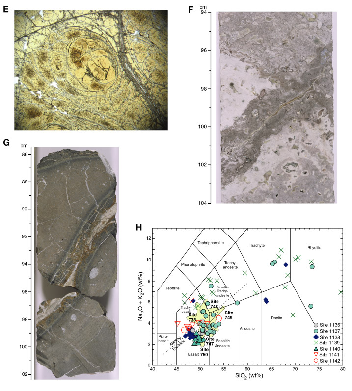
1Le Bas, M.J., Le Maitre, R.W., Streckeisen, A., and Zanettin, B., 1986. A chemical classification of volcanic rocks based on the total alkali-silica diagram. J. Petrol., 27:745–750.