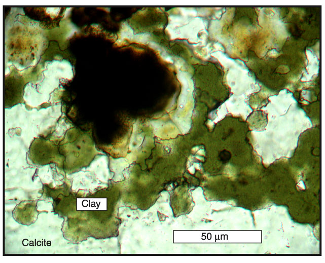
Figure F83. Photomicrograph of a thin section (from Piece 2 in Figure F82) from Sample 185-801C-27R-3, 6-11 cm. This photograph shows the calcite matrix enclosing green clay and brown iron oxides. Photomicrograph is in plane-polarized light with a 20× objective lens.
![]()