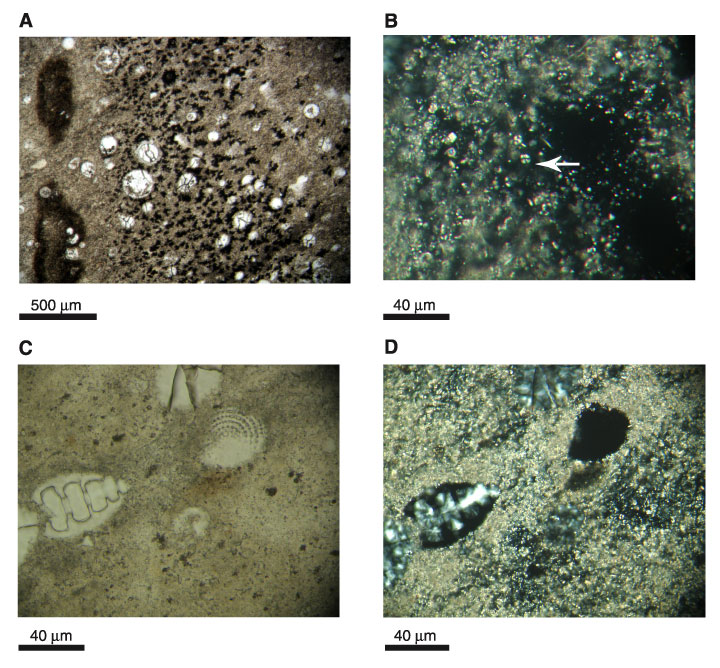
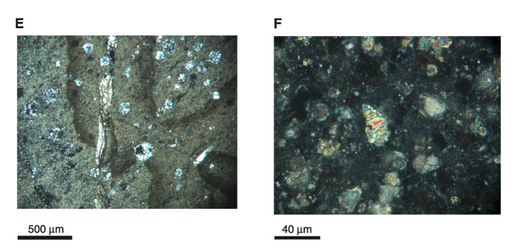
Figure F25. Photomicrographs of radiolarian chalk and radiolarian-bearing calcareous marlstone showing various stages of silica diagenesis and alteration of siliceous microfossils in radiolarian marlstones of lithologic Unit IV. A. Clear spheres are radiolarians at various stages of alteration from opal-CT to quartz (most are chalcedony). Dark patches are richer in clay minerals in a dominantly calcareous groundmass. Taken under plane-polarized light; Sample 185-1149B-18R-1, 69-71 cm (301.7 mbsf). B. Close-up view of carbonate groundmass, which largely consists of recrystallized, partially dissolved, and broken calcareous nannofossils (indicated by arrow). Taken under cross-polarized light; Sample 185-1149B-18R-1, 69-71 cm (301.7 mbsf). C. Diatom and radiolarians in calcareous matrix. Taken under plane-polarized light; Sample 185-1149B-18R-1, 69-71 cm (301.7 mbsf). D. Same field of view as in (C). Diatom to the right of the center is optically isotropic test indicating a composition of opal (opal-CT according to XRD data in Sample 185-1149B-18R-1, 9-11 cm). Taken under cross-polarized light. Radiolarian left of the center with moderately preserved morphology and optically isotropic test, whereas the infill is birefringent chalcedony. The radiolarian "ghost" partially captured at the top center of the photomicrograph shows complete conversion to chalcedony. The close juxtaposition of these different stages of diagenetic alteration suggests an overlap of the stability regimes for opal-CT and quartz. E. Calcite-filled vein intersecting thin, dark chalcedony vein. Note the halo associated with vein filling. Taken under cross-polarized light. F. Radiolarian from deeper in lithologic Unit IV (interval 185-1149B-20R-1, 52-55 cm; 321.5 mbsf) that has undergone complete recrystallization to calcite. Image taken under cross-polarized light.
![]()