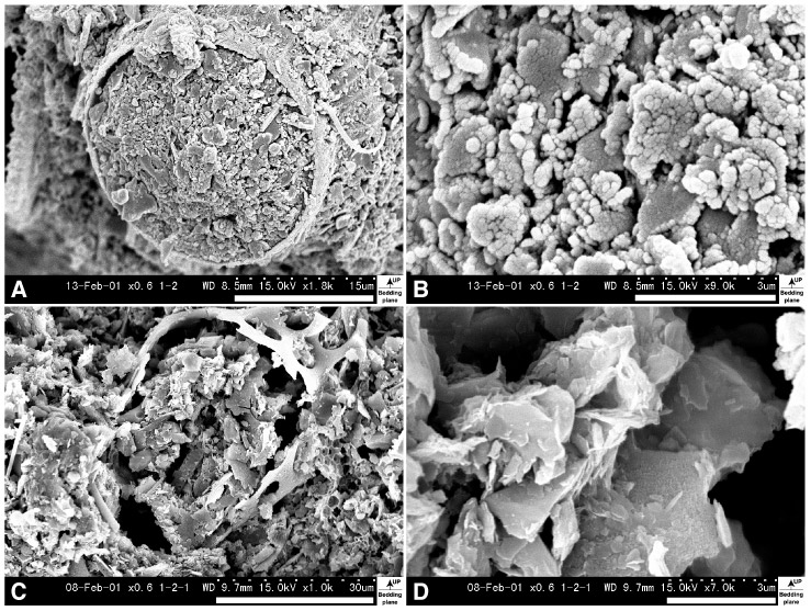
Figure F2. Comparison between both microfabrics treated by the freeze-drying and air-drying methods. Vertical views, secondary electron images at 2.13 mbsf (Sample 185-1149A-1H-2, 63-65 cm), Unit I. A. Microfabric treated by freeze-drying method. Note that fine sediments fill in radiolarian test. B. Close-up image of center of A. Clay platelets have EF contact. C. Microfabric treated by air-drying method, 60 oc, 24 hr. Volume shrinkage of sediment is 40.53%. Length shrinkage of sediment is 15.08% (vertical direction), 16.03% (lateral direction), and 16.60% (depth direction) in this micrograph. D. Close-up image of center of C. Most of clay platelets are in low-angle EF and FF contact.