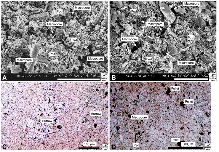
Figure F8. Microfabrics of Unit I. A. General view showing random and porous fabric from core at 2.13 mbsf (Sample 185-1149A-1H-2, 63-65 cm); vertical view, secondary electron image. B. Microfabrics from core at 52.49 mbsf (Sample 185-1149A-7H-1, 79-81 cm) showing random and porous fabric; vertical view, secondary electron image. C. Cross section from core at 2.13 mbsf (Sample 185-1149A-1H-2, 63-65 cm). Radiolarian tests in burrow are filled by fine sediments; vertical view, open nicols. D. Cross section from core at 115.60 mbsf (Sample 185-1149A-13H-5, 90-92 cm). Peds and macropores are seen in microfabric; vertical view, open
nicols.