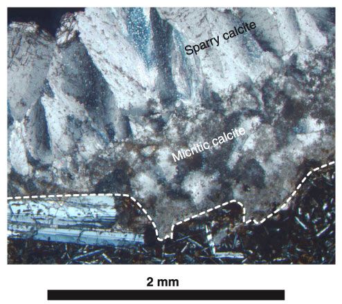
Figure
F13. Photomicrograph,
with crossed polars, of Sample 187-1155B-6R-2, 0-3 cm (see "Site
1155 Thin Sections"), showing sparry calcite growing perpendicular
to the contact with micritic calcite. The inserted white dashed line follows the
vein wall.