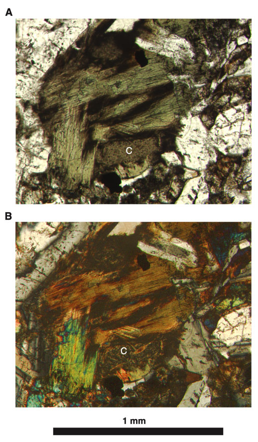
Figure F6. A. Photomicrograph in plane-polarized light of Sample 187-1162A-5R-1 (Piece 7, 25-28 cm; see "Site 1162 Thin Sections"), showing actinolite bundles (green) with chlorite (C; patchy brown/black, below and right) after clinopyroxene in metagabbro. B. Photomicrograph, with crossed polars, of the same view.
![]()