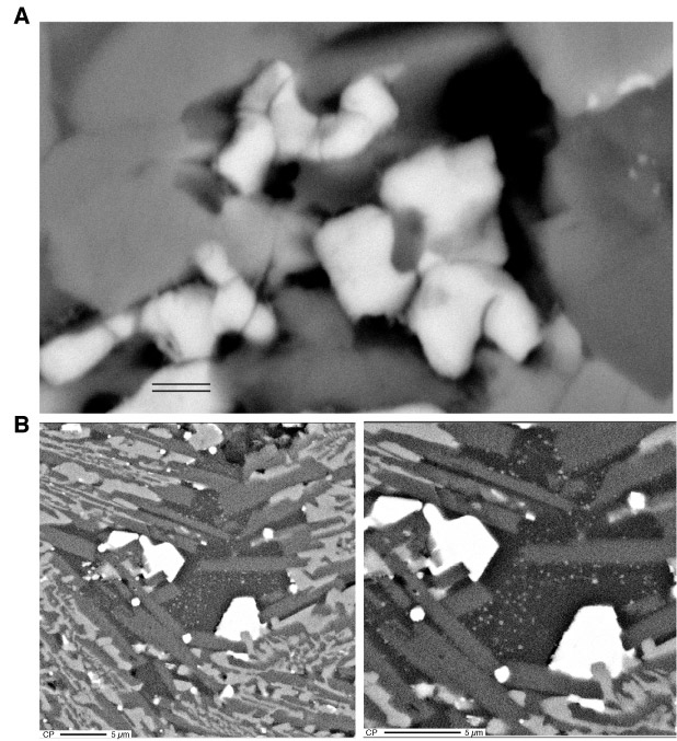
Figure F10. Scanning electron microscopic backscattered electron images showing features of the opaque minerals. A. Titanomagnetite pseudomorphs in pillow basalt show shrinkage curvature cracks. These cracks and their adjacent areas are filled or replaced by silicates as shown by the stains of darker contrast within the titanomagnetite pseudomorphs (Sample 187-1154A-8R-1, 135-140 cm [#20]). Scale bar = 1 µm. B. Submicroscopic Fe-Ti oxide mineral grains (gray-white dots) in interstitial glass (darkest area at central part of the two images) of pillow basalt (Sample 187-1159A-2R-1, 26-30 cm [#57]). The right image is an enlarged part of the central area of the left image. Three large subhedral to euhedral titanomagnetite pseudomorphs are a few micrometers across. Lath-shaped minerals are dark gray plagioclase and gray-white clinopyroxene. Scale bar = 5 µm.