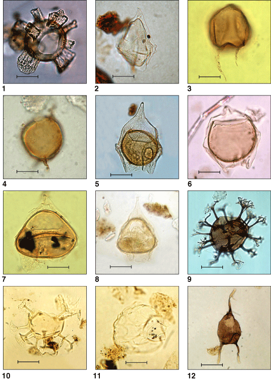
Plate P1. Illustrations of taxa, sample, and slide number. Scale bar = ~20 µm unless stated otherwise. Scanning electron microscope (SEM) photographs have varying scale bars. Many of the taxa below are also illustrated in
Brinkhuis, Sengers, et al.
(this volume). 1. Aireiana verrucosa (Sample 189-1172A-39X-5, 55-57 cm) (1). 2. Alterbidinium distinctum (Sample 189-1172A-39X-4, 85-87 cm) (2). 3, 4. Brigantedinium? sp.; (3) Sample 189-1172A-39X-3, 122-124 cm (2) (scale bar = ~15 µm); note periphragm delineating long slender apical and antapical horns. (4) Sample 189-1172A-39X-3, 128-130 cm (2) (scale bar = ~15 µm); note periphragm delineating long slender antapical horns. 5. Deflandrea antarctica group (Sample 189-1172A-39X-6, 85-87 cm) (1). 6. Deflandrea phosphoritica group (Sample 189-1172A-39X-4, 10-12 cm) (1). 7. Deflandrea sp. A (Sample 189-1170D-7R-2, 85-87 cm) (1). Note triangular endophragm. 8. Deflandrea sp. A (Sample 189-1172A-39X-4, 74-76 cm) (1). Note triangular endophragm. 9. Enneadocysta partridgei (Sample 189-1172A-39X-5, 105-107 cm) (1). 10. Enneadocysta sp. A (Sample 189-1170D-7R-1, 25-27 cm) (1); note the two distally connected antapical processes. 11. Gelatia inflata (Sample 189-1171D-4R-2, 55-57 cm) (1). 12. Octodinium askiniae (Sample 189-1171D-4R-2, 55-57 cm) (1).