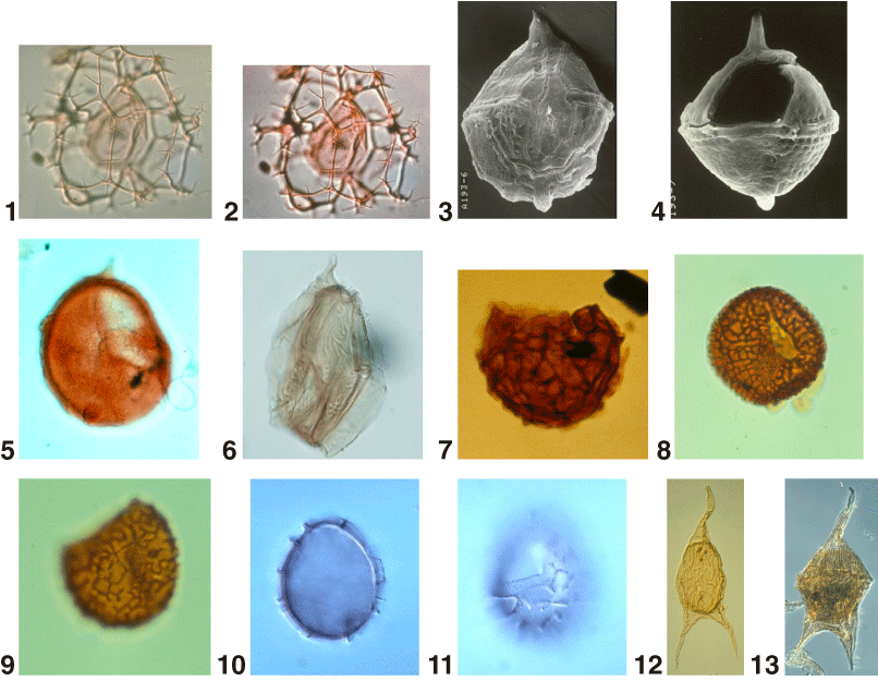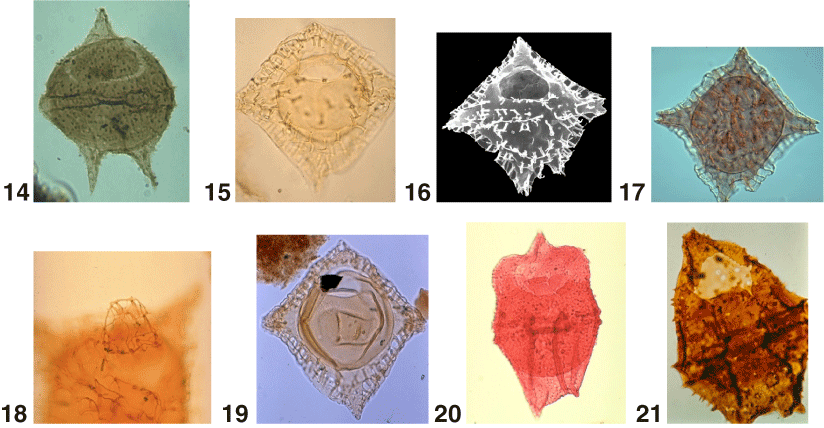
Plate P13. 1, 2. Cannosphaeropsis utinensis O. Wetzel 1933b. Same specimen (350x). 3–5. Carpatella cornuta Grigorovich 1969a. Courtesy of S.P. Damassa; (3) scanning electron micrograph (SEM), ventral surface (450x); (4) SEM, dorsal surface (450x); (5) light micrograph, optical section (450x). 6. Carpodinium obliquicostatum Cookson and Hughes 1964. 550x. 7. Cassiculosphaeridia reticulata Davey 1969a. Courtesy of J.P. Verdier. 750x. 8, 9. Cerebrocysta bartonensis Bujak in Bujak et al. 1980; (8) dorsal surface (900x); (9) right lateral view (900x). 10, 11. Cerebrocysta poulsenii de Verteuil and Norris 1996a; (10) optical section of the holotype (750x); (11) ventral view of dorsal surface of the holotype (750x), courtesy of L. de Verteuil. 12, 13. Cerodinium diebelii (Alberti 1959b) Lentin and Williams 1987. 250x. 14. Cerodinium wardenense (Williams and Downie 1966c) Lentin and Williams 1987. Dorsal surface (650x). 15, 16. Charlesdowniea columna (Michoux 1988) Lentin and Vozzhennikova 1990; (15) dorsal view of dorsal surface (350x); (16) SEM, dorsal surface (350x). 17, 18. Charlesdowniea crassiramosa (Williams and Downie 1966b) Lentin and Vozzhennikova 1989; (17) dorsal view of dorsal surface (700x); (18) operculum (350x). 19. Charlesdowniea edwardsii (Wilson 1967c) Lentin and Vozzhennikova 1989. Ventral view of dorsal surface (400x). 20. Chatangiella verrucosa (Manum 1963) Lentin and Williams 1976. Dorsal view of dorsal surface (300x). 21. Chichaouadinium vestitum (Brideaux 1971) Bujak and Davies 1983. Ventral view of dorsal surface (725x). Holotype. Courtesy of W.W. Brideaux.


![]()