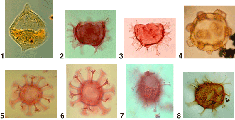
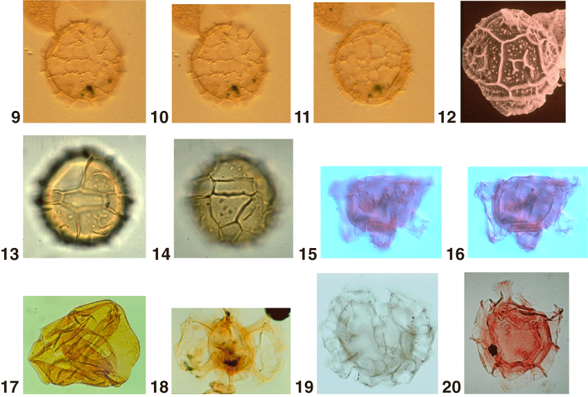
Plate P14. 1. Chichaouadinium vestitum (Brideaux 1971) Bujak and Davies 1983. Dorsal view of dorsal surface (600x). 2, 3. Chiropteridium galea (Maier 1959) Sarjeant 1983; (2) ventral view, optical section (325x); (3) dorsal view of ventral surface (325x). 4. Conosphaeridium striatoconum (Deflandre and Cookson 1955) Cookson and Eisenack 1969. Oblique antapical surface (550x). 5, 6. Cordosphaeridium cantharellus (Brosius 1963) Gocht 1969. Courtesy of S.P. Damassa; (5) optical section (350x); (6) dorsal surface (350x). 7, 8. Cordosphaeridium funiculatum Morgenroth 1966a; (7) dorsal view of dorsal surface, courtesy of S.P. Damassa. Both 400x. 9–11. Corrudinium harlandii Matsuoka 1983b. Holotype; (9) dorsal view (650x); (10, 11) optical sections (650x), courtesy of K. Matsuoka. 12–14. Corrudinium incompositum (Drugg 1970b) Stover and Evitt 1978; (12) scanning electron micrograph (SEM), oblique apical view (750x); (13) right lateral surface (750x); (14) left lateral surface (750x). 15–17. Cousteaudinium aubryae de Verteuil and Norris 1996a; (15) left ventro-lateral view of left ventro-lateral surface (450x); (16) left ventro-lateral view in optical section (450x). Figures 15, 16 courtesy of L. de Verteuil. 18. Cyclapophysis monmouthensis Benson 1976 (300x). 19. Cyclonephelium filoreticulatum (Slimani 1994) Prince et al., 1999. 450x. 20. Cyclonephelium membraniphorum Cookson and Eisenack 1962b. 300x.