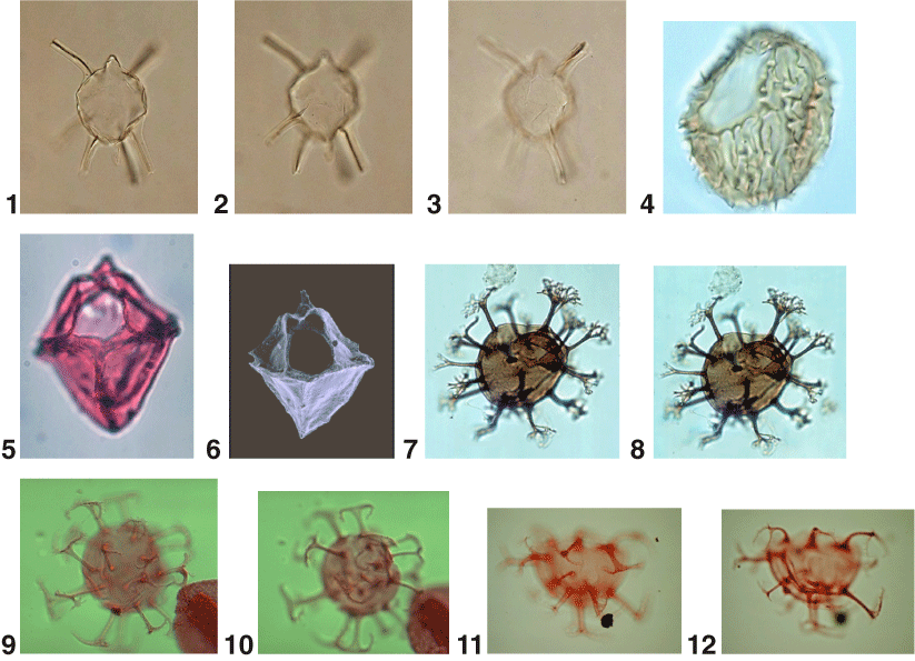
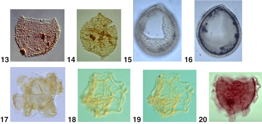
Plate P17. 1–3. Edwardsiella sexispinosa Versteegh and Zevenboom in Versteegh 1995; (1, 2) optical sections (300x); (3) dorsal surface (300x), courtesy of G.J.M. Versteegh. 4. Ellipsodinium rugulosum Clarke and Verdier 1967. Right lateral surface (950x). 5, 6. Endoscrinium campanula (Gocht 1959) Vozzhennikova 1967; (5) dorsal surface (350x); (6) scanning electron micrograph (SEM), dorsal surface (300x), courtesy of E.J. Kidson. 7, 8. Enneadocysta partridgei Stover and Williams 1995; (8) same specimen as 7 showing operculum inside. Both (300x). 9–12. Enneadocysta pectiniformis (Gerlach 1961) Stover and Williams 1995; (9, 10). same specimen (500x); (11) dorsal surface (500x); (12) same specimen as 11, ventral surface (500x). 13, 14. Epelidosphaeridia spinosa Cookson and Hughes 1964 ex Davey 1969a; (13) dorsal view of dorsal surface (700x); (14) ventral view (550x). 15, 16. Filisphaera filifera Bujak 1984; (15) dorsal surface (650x); (16) optical section (650x). 17. Florentinia mayii Kirsch 1991. 600x. 18, 19. Galeacysta etrusca Corradini and Biffi 1988. Holotype; (18) ventral view of dorsal surface (400x); (19) ventral view of ventral surface (400x). 20. Gerdiocysta conopeum Liengjarern et al. 1980. 400x.