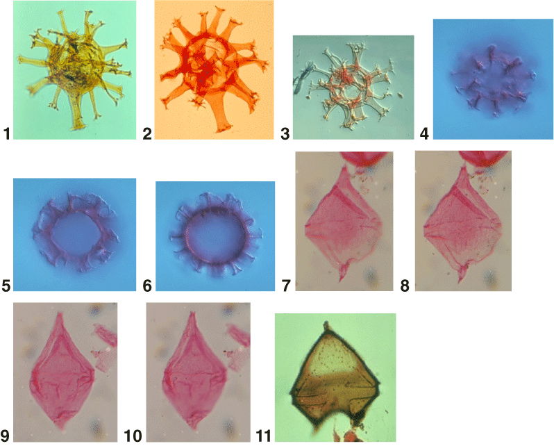
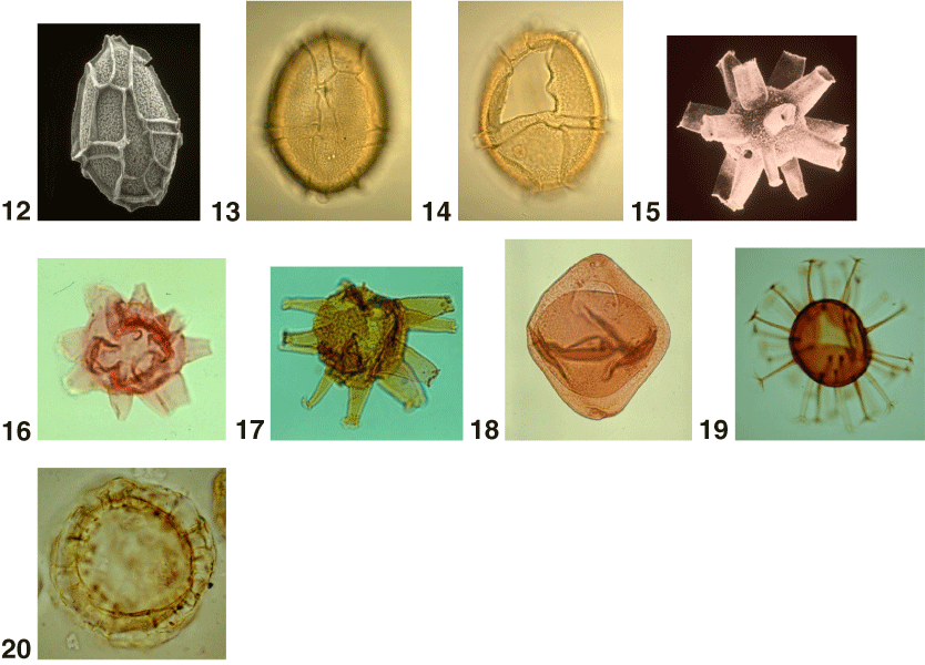
Plate P20. 1, 2. Kleithriasphaeridium loffrense Davey and Verdier 1976. Both 400x. 3. Kleithriasphaeridium readii (Davey and Williams 1966b) Davey and Verdier 1976. 300x. 4–6. Labyrinthodinium truncatum Piasecki 1980; (4) antapical surface; (6) archeopyle margin in focus. Courtesy of L. de Verteuil. All 800x. 7–10. Laciniadinium biconiculum McIntyre 1975; (7) optical section in dorsal view (500x); (8) same specimen as 7, dorsal view (500x); (9, 10). Same specimen in dorsal view (500x). 11. Lentinia serrata Bujak in Bujak et al. 1980. Optical section of holotype (650x). 12–14. Leptodinium italicum Biffi and Manum 1988; (12) scanning electron micrograph (SEM), ventral surface (625x); (13) dorsal view of ventral surface (625x); (14) same specimen as 13, dorsal view of dorsal surface (625x). 15–17. Litosphaeridium siphonophorum (Cookson and Eisenack 1958) Davey and Williams 1966b; (15) SEM (450x), courtesy of E.J. Kidson; (16) 450x; (17) 500x. 18. Manumiella seelandica (Lange 1969) Bujak and Davies 1983. 425x. 19. Melitasphaeridium pseudorecurvatum (Morgenroth 1966a) Bujak et al. 1980. Optical section (525x). 20. Membranilarnacia? picena Biffi and Manum 1988. Optical section (675x).