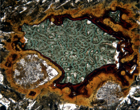
Figure F60. Photomicrograph, at higher magnification, of Sample 192-1185B-20R-1 (Piece 4, 27-29 cm) showing the large miarolitic cavity in Figure F59 (top center) (field of view = 1.4 mm; plane-polarized light; photomicrograph ID# 1185B_216).