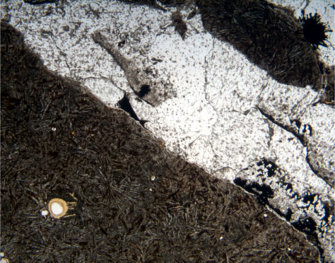
Figure F62. A 3- to 5-mm-thick vein made up of the following sequence of secondary minerals in Sample 192-1185B-5R-8 (Piece 3, 31-33 cm) (from vein walls to center): a thin (~50-100 µm thick) tan smectite edge, botryoidal concretions of an opaque mineral (probably a Mn oxyhydroxide), discontinuous "screens" consisting of tan zeolites with opaque linings, and sparry calcite. Calcite forms ~85% of the vein. The zeolite screens are fragments of the wall lining plucked away from the walls during reopening of the vein when calcite precipitated. Note a vesicle lined with zoned, yellowish tan smectite (bottom left) (field of view = 5.5 mm; plane-polarized light; photomicrograph ID# 1185B_202).
![]()