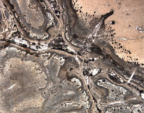
Figure F23. Photomicrograph in plane-polarized light of anhydrite-pyrite vein with pyrite surrounding anhydrite. Note the fine-grained pyrite at the outer fringes of the gray halo of silica around the veins (Section 193-1188A-7R-2 [Piece 2, 39-41 cm]; width of view = 1.38 mm. Photomicrograph ID# 1188A_40, thin section 6).
![]()