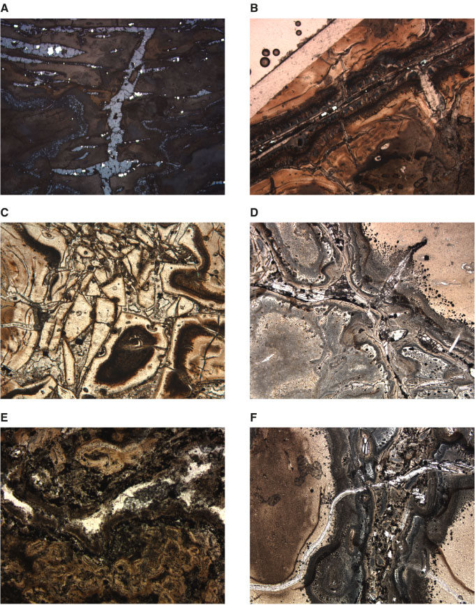
Figure F101. Photomicrographs of vein textures, Hole 1188A. A. Silica-pyrite vein cutting altered volcanic rock with offshoots parallel to flow banding (Sample 193-1188A-9R-1 [Piece 7, 68-70 cm] in reflected light; width of view = 2.75 mm. Photomicrograph ID# 1188A_57; thin section 9). B. Anhydrite-pyrite vein with cristobalite halos demonstrating two episodes of opening. Note the vein offshoot crosscutting the first halo in the upper right of the photo (Sample 193-1188A-9R-1 [Piece 7, 68-70 cm] in plane-polarized transmitted light; width of view = 2.75 mm. Photomicrograph ID# 1188A_16; thin section 9). C. Fragments of strongly silicified volcanic rock in a cristobalite-pyrite vein. Note the open-space growth on the fragments (Sample 193-1188A-9R-1 [Piece 7, 68-70 cm] in plane-polarized transmitted light; width of view = 2.75 mm. Photomicrograph ID# 1188A_45; thin section 9). D. Anhydrite-pyrite vein with pyrite surrounding anhydrite. Note the fine-grained pyrite at the outer fringes of the gray halo of silica around the veins (Sample 193-1188A-7R-2 [Piece 2, 39-41 cm] in plane-polarized transmitted light; width of view = 1.40 mm. Photomicrograph ID# 1188A_40; thin section 6). E. Quartz vein rimmed by brown clay-cristobalite and farther outward by a band of hematite and rutile with minor pyrite (Sample 193-1188A-12R-1 [Piece 12, 123-124 cm] in plane-polarized transmitted light; width of view = 1.40 mm. Photomicrograph ID# 1188A_108; thin section 11). F. Anhydrite-silica-pyrite vein with pronounced silica alteration halo, crosscut by later anhydrite vein. Note the fine-grained "dusty" pyrite at the outer fringes of the silica alteration halo (Sample 193-1188A-7R-2 [Piece 2, 39-41 cm] in plane-polarized transmitted light; width of view = 1.40 mm. Photomicrograph ID# 1188A_04; thin section 6).
![]()