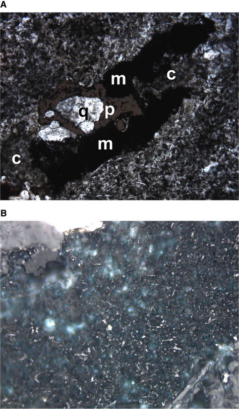
Figure F63. A. Combined plane-polarized transmitted and reflected light photomicrograph of a "magnetite" (m)-chlorite (c)-quartz (q)-pyrite (p) filled vesicle. Width of view = 1.4 mm. B. High-magnification reflected light image of the opaque portion (m) of the amygdule in (A). It consists of fine, spongy remnant magnetite in a matrix of clay (white internal reflections) and dark opaque material. Width of view = 0.275 mm (interval 193-1188F-40Z-1, 3-5 cm; Unit 65). Photomicrograph ID# 1188F_94 and 1188F_95; thin section 106).
![]()