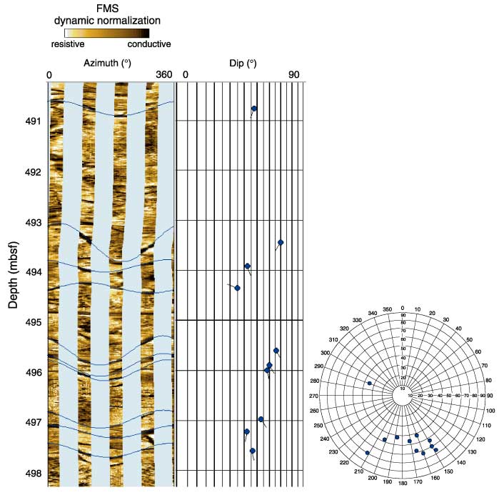
Figure F66. Formation MicroScanner (FMS) image of a fractured interval (490-498.5 mbsf) at the base of logging Unit 3. The inset displays the dip of the fractures that average 160°/60°.