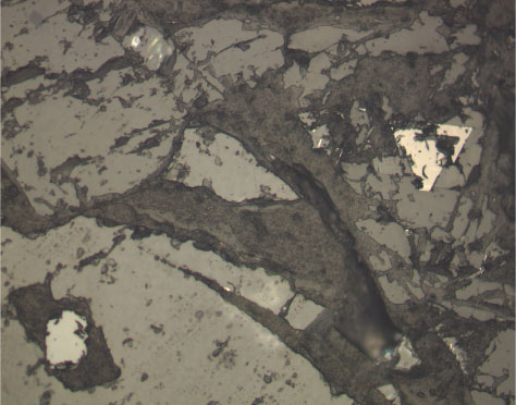
Figure F40.
Photomicrograph of a Cr spinel inclusion in olivine (bottom left) and a titanomagnetite crystal (middle right) in Unit 11 (Sample
197-1203A-32R-3 [Piece 1E, 85-87 cm]
) (reflected light; field of view = 0.5 mm; photomicrograph 1203A-120).