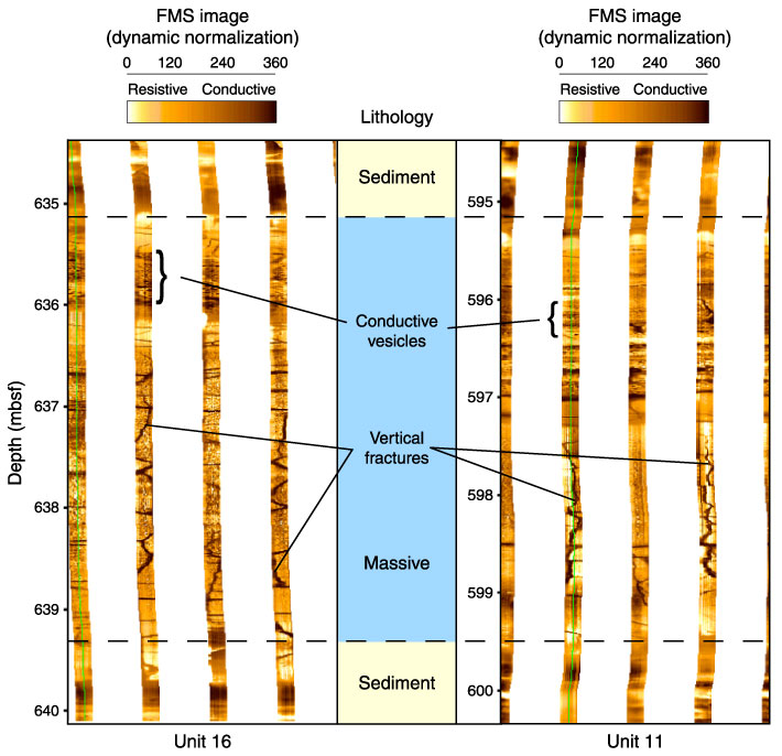
Figure F83. Detailed Formation MicroScanner (FMS) image displaying typical massive intervals within thick pahoehoe flows (Units 11 and 16). Both units present the same internal organization with horizontal vesicles on the top and vertical features in the massive part (jointing structures or pipe vesicles).