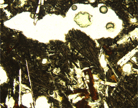
Figure F16. Photomicrograph of segregated material on margin of vesicle, with both clinopyroxene and titanomagnetite exhibiting acicular morphologies indicative of quenching (Sample 197-1204B-2R-2, 48-50 cm) (plane-polarized light; field of view = 1.25 mm; photomicrograph 1204B-157).


![]()