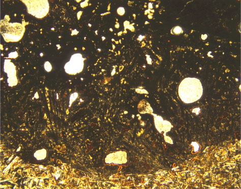
Figure F17.
Photomicrograph of a vesicle cylinder containing segregated material consisting of clinopyroxene and titanomagnetite (Sample
197-1204B-15R-1, 15-18 cm
) (plane-polarized light; field of view = 5 mm; photomicrograph 1204B-166).