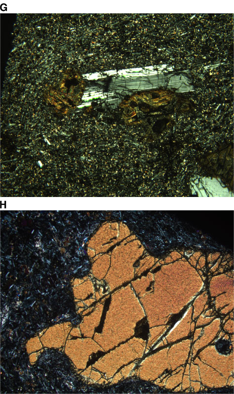
Figure F16. Photomicrographs showing plagioclase phenocrysts. G.
Plagioclase-olivine glomerocrysts (Sample 197-1205A-29R-2,
55-58 cm) (cross-polarized light; field of view = 5 mm;
photomicrograph 1205A-235). H. Highly embayed olivine
phenocryst from Unit 21 (Sample 197-1205A-37R-5,
28-29 cm) (cross-polarized light; field of view = 5 mm;
photomicrograph 1205A-257).