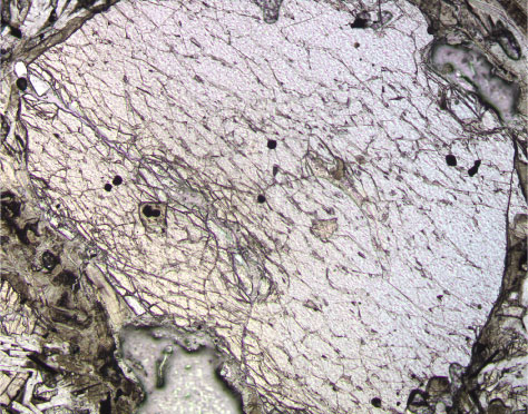
Figure F17.
Photomicrograph of olivine phenocryst with Cr spinel inclusions (dark spots) in Unit 1 (Sample
197-1206A-4R-3 [Piece 4A, 72-74 cm]
) (cross-polarized light; field of view = 1.25 mm; photomicrograph 1206A-295).